Certain Retinal Vessel Traits May Signal Balance Impairment - Review of Optometry
This story deserves to be shared
Send it to someone who'd care, or post it on your networks.

In aging adults, reduced balance is associated with fall-related injuries, nursing home admission and an increased risk of death. A new study in Investigative Ophthalmology & Visual Science found that certain measurable features of the retinal vasculature were linked to a higher likelihood of worsening balance, suggesting that retinal imaging may eventually help flag patients who could benefit from earlier intervention. The study drew on data from the Canadian Longitudinal Study on Aging, a large population-based cohort of older adults. Participants underwent retinal imaging and were followed over three years for changes in balance status. The researchers evaluated retinal vessel traits using digital fundus photographs and assessed whether those baseline characteristics predicted the later development of balance impairment, defined as the inability to stand on one leg unassisted for one minute. According to the authors, this study is the first to assess this relationship.
The researchers used a deep learning algorithm called the Quantitative Analysis of Retinal Vessel Topology and Size (QUARTZ) to measure retinal vessel morphology.
This article is an excerpt. To read the full content, visit the original source. Review of Optometry
Related articles

Eye scans link retinal thinning to slower cognitive processing in MS - Multiple Sclerosis News Today
Study highlights tests as possible tool for monitoring neurodegeneration Thinning of a layer of nerve cells in the eye's retina was associated with slower information processing in people with multiple sclerosis, independent of physical disability, a study found.
Vascular Contributions to Cognitive Impairment and Brain Health - professional.heart.org
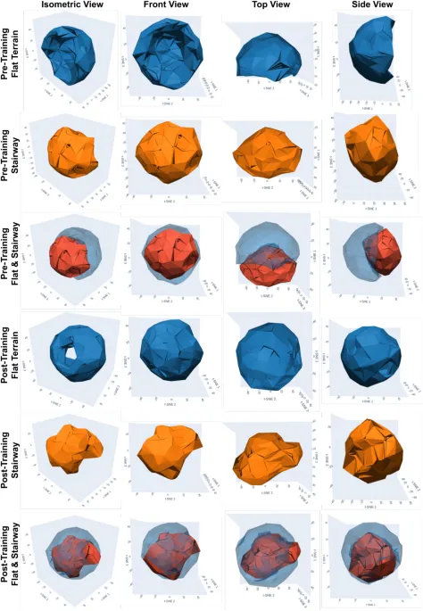
A semantic-conditioned multi-modal behavior cloning framework for terrain-specific behavior isolation in humanoid locomotion - Nature
Held-out test subset of \(\mathscr {D}\) \(\textbf{e}_{l_t} \in \mathbb {R}^{8}\)
Associations of serum phosphate levels and intravenous phosphate exposure with mortality in patients with sepsis: a multi-database retrospective study - Nature
We’re sharing this article early to provide faster access to peer-reviewed, accepted research. It is citable and carries a permanent DOI. This version is subject to further edits and will be replaced automatically by the final Version of Record. All legal disclaimers apply. AbstractAbnormal phosphate concentrations are common in sepsis, but the prognostic relevance of serum phosphate and the association between recorded intravenous phosphate exposure and mortality remain uncertain. This retrospective cohort study used MIMIC-III, MIMIC-IV, and a single-center database from the First Affiliated Hospital of Chongqing Medical University. Adults with ICD-coded sepsis, a first ICU admission, and an ICU stay longer than 24 h were included. Admission phosphate was evaluated with restricted cubic spline regression for 28- and 90-day mortality. Recorded intravenous phosphate exposure was assessed using multivariable logistic regression in the 48- and 72-h landmark populations. Propensity-score matching and inverse-probability-of-treatment weighting were performed at both landmarks. A target-trial emulation was also performed. Cohorts 1 and 2 included 15,426 and 967 patients, respectively.
Comparative evaluation of unilateral anterior crossbite and monoiodoacetate injection models for temporomandibular joint osteoarthritis in rats - Nature
We’re sharing this article early to provide faster access to peer-reviewed, accepted research. It is citable and carries a permanent DOI. This version is subject to further edits and will be replaced automatically by the final Version of Record. All legal disclaimers apply. AbstractTo systematically compare unilateral anterior crossbite (UAC) and monosodium iodoacetate (MIA) models for temporomandibular joint osteoarthritis (TMJOA) research. This study used healthy adult male SPF-grade Sprague–Dawley (SD) rats, aged 6–8 weeks and weighing 180–200 g, with a total of 24 rats. After a 7-day acclimatization period, the rats were randomly and equally allocated into three groups: a normal control group (NC), an occlusion intervention group (UAC), and a drug injection group (MIA). Condylar height at the anterior, central, and posterior regions was quantitatively measured via a computer-aided image analysis system to elucidate the observed morphological alterations. Pathological assessments of the TMJ were conducted using Safranin O-Fast Green staining, hematoxylin and eosin (HE) staining, and immunohistochemistry (IHC).
0 comments
No comments yet. Be the first to share your thoughts!