Retinal calcium waves coordinate uniform tissue patterning of the Drosophila eye - Science | AAAS
This story deserves to be shared
Send it to someone who'd care, or post it on your networks.
This article is an excerpt. To read the full content, visit the original source. Science | AAAS
Related articles
Author Correction: Ecdysone signaling-induced dumpless1 expression controls nurse cell dumping in Drosophila oogenesis - Nature
volume 17, Article number: 10957 (2026) Cite this article Correction to: Nature Communications https://doi.org/10.1038/s41467-025-63973-3, published online 7 October 2025Following the publication of this article, we identified an error in the Source Data file accompanying the paper. In the worksheet for Fig. 8o, both the control and the integrin βPS RNAi experimental group data columns were incorrectly pasted during data compilation—they were inadvertently populated with a subset of measurements (52 values each) that had been extracted from the 7-day samples of the CG12942 overexpression experiment (Fig. 3n), rather than the correct data from the independent integrin βPS knockdown rescue experiment. We have now retrieved the original experimental images and measurement records for the integrin βPS RNAi experiment (including both control and integrin βPS RNAi groups). Reanalysis using the correct data confirms that the conclusion remains unchanged: knockdown of integrin βPS in SFCs partially rescues the egg length defects in dumpless1 mutant females. The Source Data file and the scatter plot for Fig.
Nuclear adaptation to intracellular mechanical forces regulates the phagocytic capacity of Drosophila macrophages in vivo - Nature
We’re sharing this article early to provide faster access to peer-reviewed, accepted research. It is citable and carries a permanent DOI. This version is subject to further edits and will be replaced automatically by the final Version of Record. All legal disclaimers apply. AbstractHow macrophages fine-tune their appetite to fulfil their clearance role is incompletely understood. Phagocytosis exacerbates cytoplasmic crowding, but whether macrophages can sense this increase and use it to regulate their phagocytic capacity has never been investigated. Here, we test the hypothesis that phagosomes exert a strain on the nucleus, which in turn responds to the intracellular mechanical inputs influencing macrophage appetite. Combining genetic manipulation and live in vivo imaging, we show that engulfed apoptotic bodies indent and deform the nucleus of Drosophila embryonic macrophages, and that phagocytosis increases Lamin B levels. Importantly, we show that silencing Lamin B or components of the LINC complex negatively regulates apoptotic body engulfment in vivo.
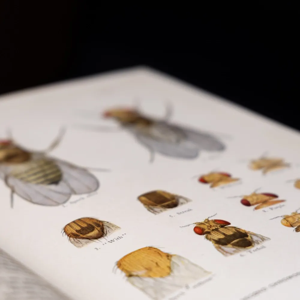
Object 24 | Drosophila Illustrations - Carnegie Science
In the spring of 1910, Thomas Hunt Morgan peered into one of the hundreds of glass milk bottles that filled his cramped and famously messy laboratory at Columbia University and spotted something he had never before seen. Among the thousands of red-eyed fruit flies he had been breeding for nearly a year was a single male with white eyes. That one small fly was the first in a series of discoveries by Morgan and his team that would transform the study of heredity. Over the following decades, Morgan's "fly group" showed that genes are physical units lined up in a fixed order along chromosomes and that their positions could be mapped, laying the foundation of modern genetics. Carnegie Science made much of this research possible, providing sustained funding to the group from 1915 until 1945 and publishing its findings in a series of volumes, which were accompanied by exquisitely detailed ink drawings and watercolors of fruit flies by Edith Wallace, Morgan's technician and the team's artist. Her illustrations, representing the fly group’s work, are Object 24 in our #Carnegie125 series. Before the Flies Morgan was born into a prominent Kentucky family in 1866. After earning his Ph. D.

Scientists find a hidden retinal defense against vision loss - Science Daily
Many diseases that lead to vision loss have something important in common: they gradually damage the retina, the layer of light-sensitive tissue at the back of the eye that makes sight possible. Scientists understand many of the physical changes that occur as retinal degeneration progresses, but they know far less about the chemical signals that influence how the retina reacts to injury. Researchers at Scripps Research, working with scientists at UC San Diego and the Lowy Medical Research Institute, have now identified a naturally occurring molecule called erucamide that appears to help coordinate communication between cells in the retina. In a study published in Nature Neuroscience on June 19, 2026, the team found that erucamide levels decline as photoreceptors, the light-sensing cells responsible for detecting visual information, begin to die.
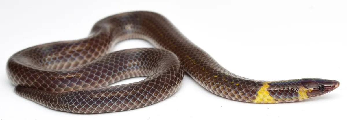
Rolling without wheels: How dwarf reedsnakes coordinate their bodies for high-speed escapes - Phys.org
A species of dwarf reedsnake, Pseudorabdion saravacense, investigated in the study. Credit: Yosuke Kojima Small snakes called dwarf reedsnakes can escape danger by rolling rapidly along the ground. Researchers investigated how they perform this unusual form of locomotion using multi-angle video analyses. The results show that the snakes use precisely coordinated body movements that keep their heads facing the direction of travel and at a relatively stable height, even as the posterior body undergoes large rotational movements.
0 comments
No comments yet. Be the first to share your thoughts!