An iron compass in the liver: superparamagnetic macrophages give homing pigeons a magnetic sense - Nature
This story deserves to be shared
Send it to someone who'd care, or post it on your networks.
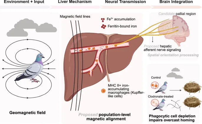
In a recent study published in Science, Lisowski et al. identified superparamagnetic, iron-loaded macrophages in the pigeon liver and showed that depletion of clodronate-sensitive phagocytic cells impaired homing under overcast skies, when solar cues were unavailable.1 Together, these observations point to a light-independent magnetoreceptive system outside the head and broaden current models of magnetoreception beyond receptors based on cryptochrome, magnetite, and ion channels in the eye and beak.2Magnetic measurements pointed the way (Fig. 1). Vibrating-sample magnetometry revealed a magnetic blocking transition near 12 K in liver and spleen, the signature of superparamagnetic nanoparticles, absent from muscle and beak. The measured signal could not be explained by isolated paramagnetic ferric iron, and low-temperature hysteresis indicated long-range magnetic order: not merely iron in the liver, but a biological nanomagnet. Prussian blue staining localized ferritin-bound iron to MHC II+ cells in hepatic sinusoids, abundant in liver, sparse in spleen, and absent from brain, eye, muscle, and beak. Fig. 1A liver-based route for magnetic orientation in homing pigeons.
This article is an excerpt. To read the full content, visit the original source. Nature
Related articles
Nuclear adaptation to intracellular mechanical forces regulates the phagocytic capacity of Drosophila macrophages in vivo - Nature
We’re sharing this article early to provide faster access to peer-reviewed, accepted research. It is citable and carries a permanent DOI. This version is subject to further edits and will be replaced automatically by the final Version of Record. All legal disclaimers apply. AbstractHow macrophages fine-tune their appetite to fulfil their clearance role is incompletely understood. Phagocytosis exacerbates cytoplasmic crowding, but whether macrophages can sense this increase and use it to regulate their phagocytic capacity has never been investigated. Here, we test the hypothesis that phagosomes exert a strain on the nucleus, which in turn responds to the intracellular mechanical inputs influencing macrophage appetite. Combining genetic manipulation and live in vivo imaging, we show that engulfed apoptotic bodies indent and deform the nucleus of Drosophila embryonic macrophages, and that phagocytosis increases Lamin B levels. Importantly, we show that silencing Lamin B or components of the LINC complex negatively regulates apoptotic body engulfment in vivo.

Found: Oldest Evidence for an Organismal Magnetic Compass - Science and Culture Today
Research conducted by a group led by scientists from Cambridge University and the Max Planck Institute has found the oldest evidence for the possible use of a magnetic compass by an organism.1 The evidence exists in so-called magnetofossils found in marine sediments that have been dated to 97 million years old. The material in the fossils is magnetite, which is one of the theorized mechanisms for animal magnetoreception. Magnetite (Fe3O4) is an iron oxide usually found as single domain crystals approximately 20-50 nanometers in diameter, making it challenging to detect in organisms. The crystals are permanent magnets that align with Earth’s magnetic field. Several animal species have been found to form biogenic magnetite, including magnetotactic bacteria and some species of fish, including salmon and trout.2 There is also some evidence that a number of genes associated with the formation of magnetite in organisms (biomineralization) are shared by bacteria and eukaryotic (multicellular) animals.3 Bacteria use the compass to guide their movement in finding favorable conditions.
How one cell becomes a body: DNA ‘typewriter’ reveals developmental history of mouse embryo - BioTechniques
A DNA ‘typewriter’ records the history of 1.3 million cells in an early mouse embryo, telling the tale of mammalian development from fertilization to organ formation like never before. Researchers from the University of Washington School of Medicine (WA, USA) have used a molecular tracking technology called DNA Typewriter to record the cellular lineage of a mouse embryo over nearly 2 weeks of development, from a single cell to over a million cells, with the potential to shed light on the origins of a range of congenital defects, neurodevelopmental disorders, genetic conditions and cancers.
0 comments
No comments yet. Be the first to share your thoughts!