Red-sensitive pigments in mantis shrimp eyes act as reversible light switches - Phys.org
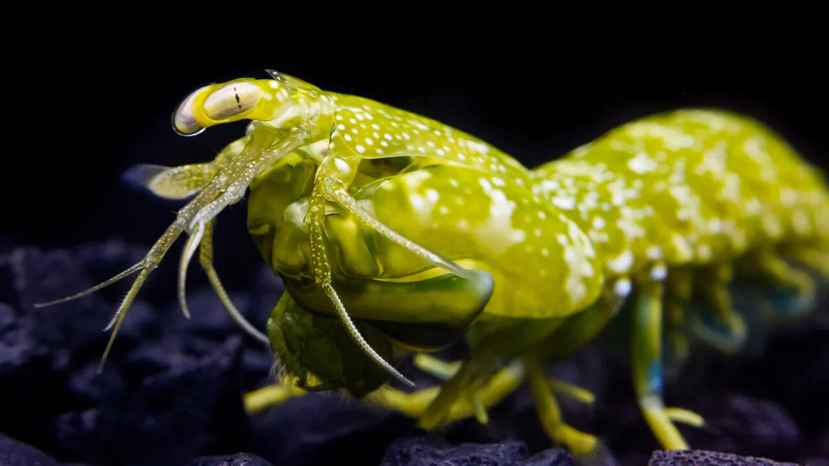
With about 30 visual pigments, compared to just three in humans, mantis shrimp possess a visual system that remains a mystery. Researchers from ULiège and Humboldt University in Berlin have, for the first time, analyzed these pigments one by one and discovered that some detect red light that very few animals can perceive. This finding, published in Science Advances, has potential applications in optogenetics.
Vision is one of the most sophisticated sensory systems; it relies on proteins (opsins) that capture light and convert the signal into an electrical message that the brain can interpret. When combined with retinal, a small, universal molecule derived from vitamin A, opsins form the visual pigments (rhodopsins) that absorb light in the photoreceptors of the retina. In humans, three cone pigments are sufficient for color perception.
This article is an excerpt. To read the full content, visit the original source. Phys.org
Related articles
Mapping genetic switches behind rice seedlings’ cold response | Newswise - Newswise
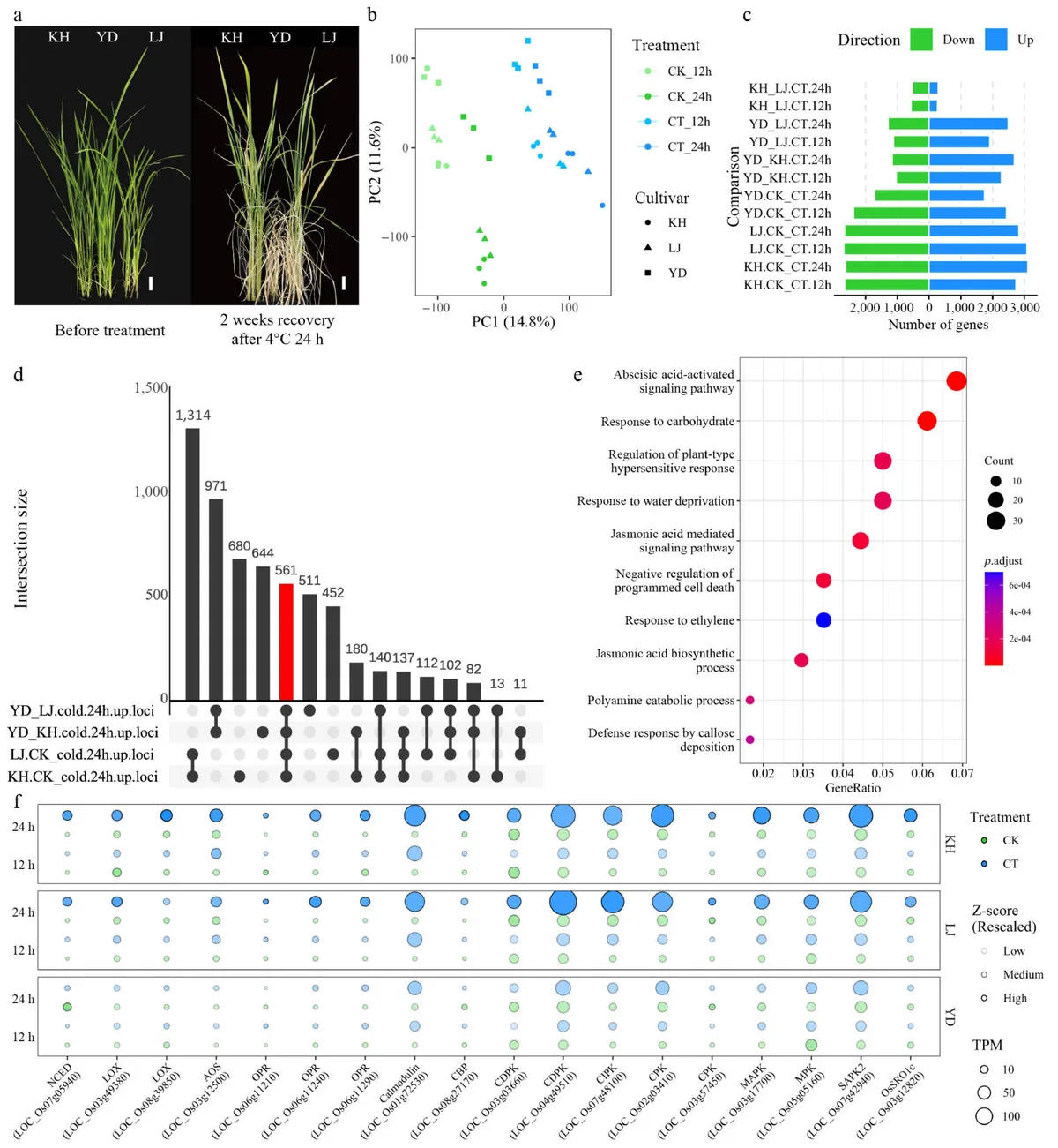
Rice originated in subtropical environments, yet its cultivation has expanded into regions where low temperatures threaten growth, development, and yield. Japonica rice generally tolerates cold better than indica rice, and researchers have linked this adaptation to changes in stress-responsive gene expression. Much attention has focused on C-repeat binding factors, a group of transcription factors that help coordinate plant cold responses. However, these proteins may directly regulate only 10%–20% of cold-regulated genes, suggesting that many other regulators operate alongside them. For numerous rice genes, the identities of those regulators and the locations of the DNA sequences they recognize remain unclear. A wider map of these interactions is therefore needed to understand cold adaptation and identify potential targets for crop improvement.
Experimental characterization and scaling of flexural rigidity in sewn multilayer structures - Nature
We’re sharing this article early to provide faster access to peer-reviewed, accepted research. It is citable and carries a permanent DOI. This version is subject to further edits and will be replaced automatically by the final Version of Record. All legal disclaimers apply. AbstractSewing is one of the established methods used to fabricate and assemble multilayer insulation (MLI) blankets for aerospace applications, yet the resulting effective flexural behavior remains insufficiently understood. This study investigates the room-temperature, quasi-static flexural behavior of MLI blankets composed of aluminized BoPET radiation shields and polyester net spacers across structural configurations of 2, 6, 12, and 24 layers. A full factorial design was employed to evaluate the influence of the number of layers, stitch density, needle thread tension, and needle foot force. Statistical analyses demonstrated that the number of layers is the dominant factor governing flexural response. Among the sewing parameters, needle thread tension was associated with modifications in bending length, while needle foot force primarily affected local compaction.
Targeted pathway mining reveals novel modified hexapeptides from the demiguisin family - Nature
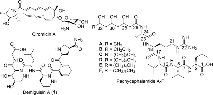
IntroductionBirds rely on a collection of behavioural, physiological, and chemical defences to mitigate the constant threat posed by pathogens and parasites in their environment1. Preen oil from the uropygial gland (UG) is a key component of avian integumentary immunity, supporting feather maintenance and waterproofing, while the UG is also known to harbour diverse microbes, including γ- and α-Pseudomonadota and Actinomycetota, that contribute to feather defense2,3. We recently exemplified a case of the bacterial isolate Amycolatopsis sp. PS_44_ISF1 derived from the UG of the toxic bird Pachycephala schlegelii that can directly contribute to the bird’s chemical defence system, by producing bioactive metabolites such as the antifungal macrolactam ciromicin A3,4, the co-secreted tensioactive lipopeptide family pachycephalamides, and a highly modified hexapeptide, named demiguisin A (1) (Fig. 1). The chemical structure of 1, caught our interest, as it contains non-proteinogenic piperazic acid (hexahydropyridazine-3-carboxylic acid, Piz) residues within the sequence Gly–β,γ-OH-Piz–Leu–Piz–DHPA–γ-c-Pro (Fig. 1)3,5.
0 comments
No comments yet. Be the first to share your thoughts!