Organism-wide cellular dynamics and epigenomic remodeling in mammalian aging - Science | AAAS
This story deserves to be shared
Send it to someone who'd care, or post it on your networks.
This article is an excerpt. To read the full content, visit the original source. Science | AAAS
Related articles
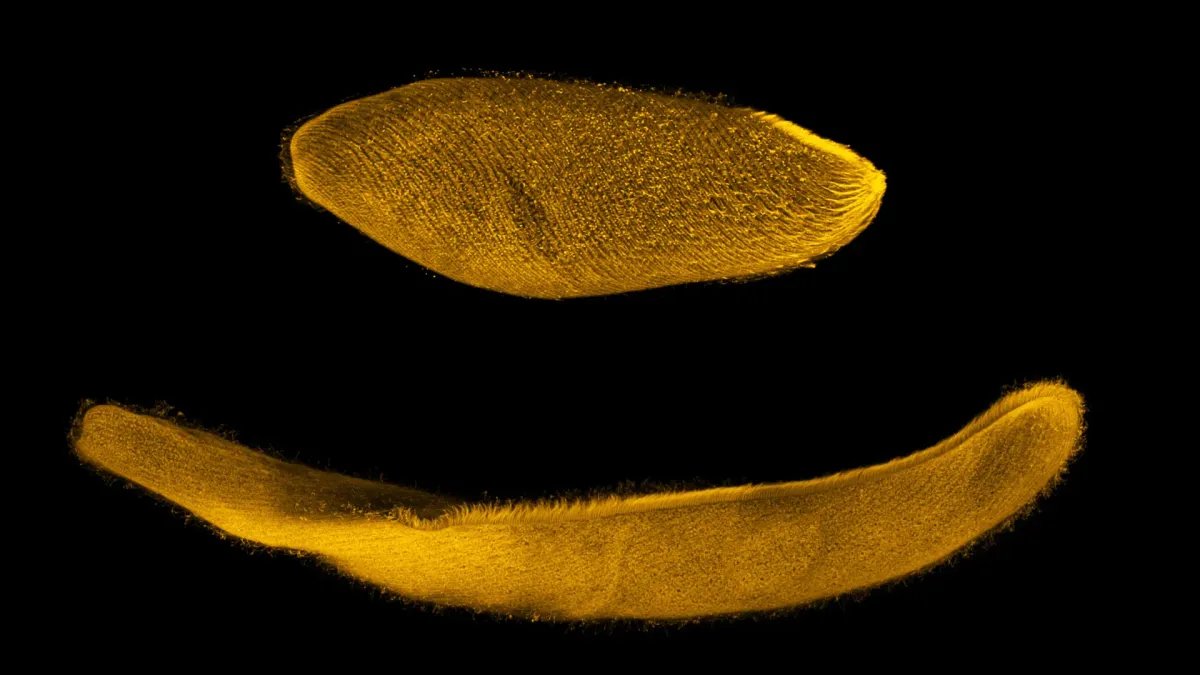
This tiny organism can shrink to one quarter its size in milliseconds - Science Daily
A tiny aquatic organism made of just one cell can compress its body to one quarter of its original length in under five milliseconds, hundreds of times faster than a person can blink. Scientists have now identified the unusual mechanism behind this remarkable movement. The organism, Spirostomum ambiguum, relies on calcium ions and a specialized protein network arranged like a fishnet. This system allows it to contract far more quickly than human muscle, and researchers say the discovery could eventually help guide the development of faster artificial muscles and synthetic cellular machinery.
The World’s Largest Living Organism Has Been Making Strange Sounds — Scientists Finally Recorded Them - Futura, le média qui explore le monde
Molecular architecture of Cryptococcus cell walls reveals species-specific chitosan-dependent remodeling - Nature
We’re sharing this article early to provide faster access to peer-reviewed, accepted research. It is citable and carries a permanent DOI. This version is subject to further edits and will be replaced automatically by the final Version of Record. All legal disclaimers apply. AbstractCryptococcus neoformans and Cryptococcus gattii are fungal pathogens that cause life-threatening infections, including cryptococcal meningitis. A distinctive feature of the cryptococcal cell wall is the extensive deacetylation of chitin to chitosan, a modification that is essential for virulence but whose structural role in cell-wall organization remains poorly understood. Here, we analyze the cell walls of wild-type strains of both species and their avirulent chitosan-deficient mutants, which serve as vaccine candidates. Loss of chitosan disrupted cell morphology and altered cell-wall ultrastructure, with more pronounced defects in C. neoformans. Solid-state NMR reveals that aggregated α-(1→3)-glucans form the principal rigid domain of the cell wall in both species and are closely associated with chitin microfibrils, whereas surrounding β-glucans and mannoproteins constitute a more dynamic matrix.
Phospholipid remodeling of the cardiomyocyte membrane affects cardiac function - Nature
AcknowledgementsWe thank Yukiko Banzai, Yuko Ishiyama, Asami Ogawa, and Yingda Xiao of the University of Tokyo for their assistance with animal experiments and laboratory work. We are grateful to Yuriko Sakamaki of the Ochanomizu Research Facility, Institute of Science Tokyo, for her technical support with electron microscopy sample preparation and imaging. FundingThis work was supported by Grants-in-Aid for Scientific Research, Japan [grant Nos. 24K19023 to S. B. and 22K08174 to H. T.]; Sakakibara Heart Foundation Cardiovascular Research Program 2023 (to S. B.); Japan Foundation for Applied Enzymology (to S. B.). I. K. was supported by a Grant-in-Aid for Scientific Research (S) of the Japan Society for the Promotion of Science and by the Japan Agency for Medical Research, Development (JP20ek0210152, JP18gm6210010, JP20ek0210141, JP20ek0109440, JP20ek0109487, JP17gm0810013, JP18km0405209, JP21ek0109543, JP21ek0109569, JP21tm0724601, JP22ama121016, JP22ek0210172, JP22ek0210167, JP22bm1123011). Author informationAuthor notesThese authors contributed equally: Satoshi Bujo, Takayuki Fujiwara, Masayuki Kubota.
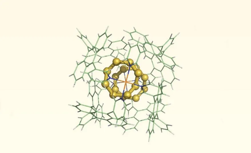
Elemental sulfur found in mammalian cells for first time - Chemistry World
© Uladzimir Barayeu et al., Science 2026 Analysis suggests that there could be tens of grams of S8 across a human body
0 comments
No comments yet. Be the first to share your thoughts!