Deep learning model automates right ventricular assessment from echocardiograms - News-Medical
This story deserves to be shared
Send it to someone who'd care, or post it on your networks.

Mostafa Al-Alusi, MD, MS, and Patrick T. Ellinor, MD, PhD, of the Mass General Brigham Heart and Vascular Institute, are the lead and senior authors of a paper published in Nature Cardiovascular Research, "Deep Learning-Enabled Assessment of Right Ventricular Size and Function is Associated with Cardiovascular Outcomes. "
Q: What challenges or unmet needs make this study important?
This article is an excerpt. To read the full content, visit the original source. News-Medical
Related articles
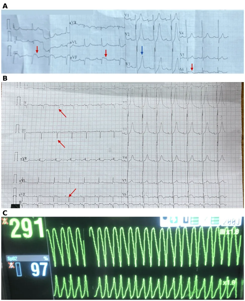
Blocking the Sympathetic Storm: Adjunctive Left Stellate Ganglion Block for Refractory Ventricular Tachycardia After Inferolateral Myocardial Infarction - Cureus
Electrical storm following myocardial infarction (MI) can be difficult to control when antiarrhythmic drugs and cardioversion fail. We describe a 53-year-old man with hypertension and previously undiagnosed diabetes who presented with an inferolateral ST-segment elevation MI caused by total occlusion of the proximal left circumflex artery, which was treated with primary percutaneous coronary intervention and drug-eluting stent placement. He then developed marked leukocytosis with a systemic inflammatory response and acute kidney injury (peak creatinine, 4.03 mg/dL), which resolved with empirical antibiotic therapy and supportive renal care. On day 6, he experienced recurrent syncope associated with sustained monomorphic ventricular tachycardia (VT) that persisted despite intravenous amiodarone and lidocaine, electrolyte correction, and repeated cardioversion, necessitating sedation and mechanical ventilation. Repeat coronary angiography demonstrated a patent stent. VT ceased following an ultrasound-guided left stellate ganglion block (SGB) using 1% lidocaine and dexamethasone, which resulted in Horner syndrome. Antiarrhythmic therapy was continued, and the patient was extubated wit
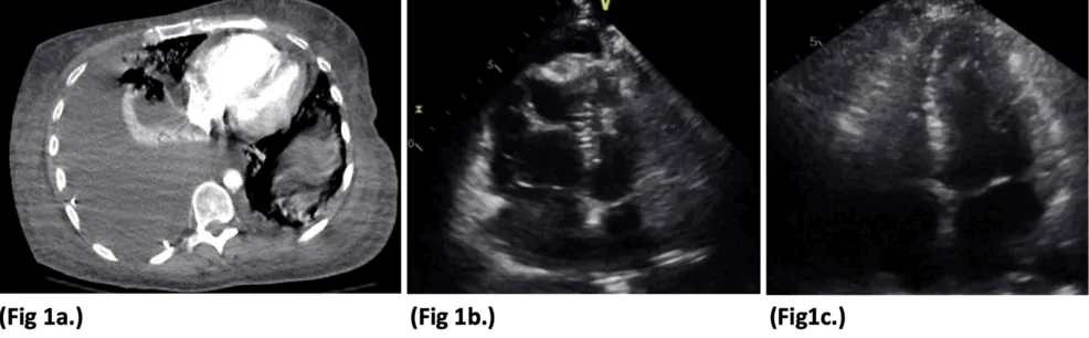
Severe Right Ventricular Dilation Due to Selenium Deficiency in a Patient After Gastric Bypass - Cureus
Selenium deficiency is a rare but reversible cause of cardiomyopathy increasingly recognized as a late nutritional complication of bariatric surgery, yet it is not consistently included in routine postoperative surveillance. This report describes a 40-year-old woman who presented three years after Roux-en-Y gastric bypass (RYGB) with progressive dyspnea, weakness, lower extremity edema, and dizziness in the setting of nonadherence to postoperative nutritional supplementation. She was hypotensive and bradycardic with jugular venous distension, elevated B-type natriuretic peptide (BNP), and chest imaging showing cardiomegaly with a right-sided pleural effusion. Echocardiography revealed severe, isolated right ventricular and atrial dilation with reduced right-sided systolic function, and further evaluation identified a markedly low serum selenium level (25 µg/L) with normal thyroid function and other trace elements. She was treated with intravenous selenium repletion (150 µg/day) alongside inotropic and diuretic support, with rapid hemodynamic improvement and complete resolution of right ventricular and atrial dilation on repeat echocardiography within one week. This case illustrates
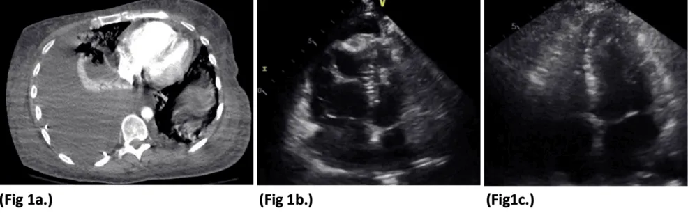
Severe Right Ventricular Dilation Due to Selenium Deficiency in a Patient After Gastric Bypass - Cureus
Selenium deficiency is a rare but reversible cause of cardiomyopathy increasingly recognized as a late nutritional complication of bariatric surgery, yet it is not consistently included in routine postoperative surveillance. This report describes a 40-year-old woman who presented three years after Roux-en-Y gastric bypass (RYGB) with progressive dyspnea, weakness, lower extremity edema, and dizziness in the setting of nonadherence to postoperative nutritional supplementation. She was hypotensive and bradycardic with jugular venous distension, elevated B-type natriuretic peptide (BNP), and chest imaging showing cardiomegaly with a right-sided pleural effusion. Echocardiography revealed severe, isolated right ventricular and atrial dilation with reduced right-sided systolic function, and further evaluation identified a markedly low serum selenium level (25 µg/L) with normal thyroid function and other trace elements. She was treated with intravenous selenium repletion (150 µg/day) alongside inotropic and diuretic support, with rapid hemodynamic improvement and complete resolution of right ventricular and atrial dilation on repeat echocardiography within one week. This case illustrates
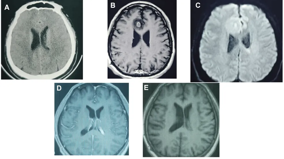
Radiologically Suspected Ventricular Infection in a Patient With Recent Self-Extraction of a Carious Tooth: A Case Report - Cureus
A brain abscess is a potentially life-threatening intracranial infection that usually requires individualized management. The possible source of infection may remain unclear when microbiological confirmation is unavailable. A 48-year-old man developed headache, fever, chills, and agitation approximately two weeks after self-extraction of a carious tooth. However, an odontogenic source was not microbiologically confirmed, and the temporal association alone did not establish causality. He also reported a history of swimming in a public pool, although the timing and characteristics of this exposure were unclear. After receiving empirical antimicrobial therapy at a local hospital without substantial improvement, he presented to Beijing Tiantan Hospital with a six-day history of fever and headache. Contrast-enhanced brain magnetic resonance imaging (MRI) showed a right frontal lesion radiologically consistent with a brain abscess and abnormalities of the left lateral ventricle and ependyma suggestive of ventriculitis, with possible intraventricular empyema. The suspected ventriculitis and intraventricular empyema were based on imaging findings and were not microbiologically confirmed. C
New clinical decision support tool aims to improve chronic kidney disease care - News-Medical
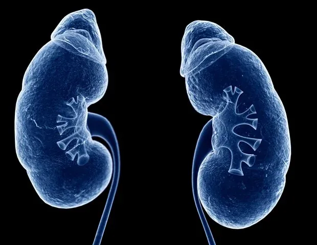
Investigators have developed and validated a clinical decision support tool to improve the diagnosis and treatment of chronic kidney disease (CKD) at the point of care. The research will be presented at ASN Kidney Week 2026 October 22–25. Called Treat CKD, the tool draws on electronic health records data to identify CKD, classify risk by Kidney Disease: Improving Global Outcomes criteria, calculate kidney failure risk using the Kidney Failure Risk Equation, estimate cardiovascular risk using American Heart Association PREVENT equations, and provide recommendations for albuminuria testing, blood pressure management, cholesterol-lowering therapy, cardio-kidney therapies, and nephrology referral.
Counseling Kidney Donors on Future Pregnancy, With Amit Garg, MD, PhD - HCPLive
Future pregnancy plans don’t preclude living kidney donation, but candidates should be counseled on gestational hypertension risk. Future pregnancy plans need not rule out living kidney donation, but candidates should be counseled on hypertensive risk. New data on pregnancy after living kidney donation give transplant teams clearer numbers to share with donor candidates who may want children in the future.¹In a matched cohort study from 3 Canadian provinces, presented at American Society of Nephrology (ASN) Kidney Week 2026 by Amit X. Garg, MD, PhD, and published simultaneously in JAMA, most donor pregnancies were uncomplicated, with a modest increase in hypertensive disorders of pregnancy.¹ Will Donating a Kidney Affect Future Pregnancies? In 2024, more than 40,000 living kidney donations were reported across 88 countries.¹ For donors who may become pregnant later, Garg said the central question is whether donation will affect those pregnancies.
0 comments
No comments yet. Be the first to share your thoughts!