Blocking the Sympathetic Storm: Adjunctive Left Stellate Ganglion Block for Refractory Ventricular Tachycardia After Inferolateral Myocardial Infarction - Cureus
This story deserves to be shared
Send it to someone who'd care, or post it on your networks.
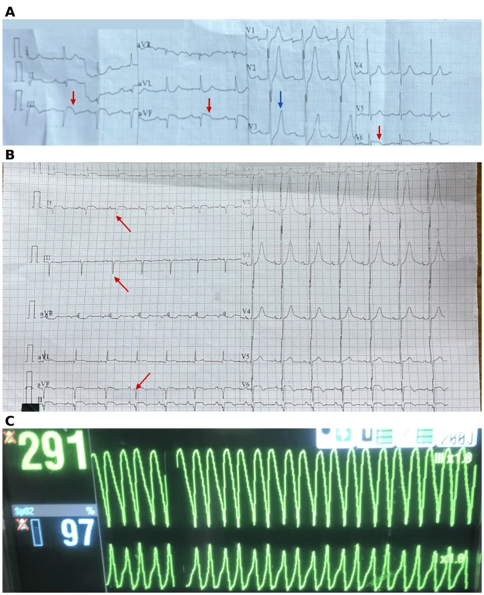
Electrical storm following myocardial infarction (MI) can be difficult to control when antiarrhythmic drugs and cardioversion fail. We describe a 53-year-old man with hypertension and previously undiagnosed diabetes who presented with an inferolateral ST-segment elevation MI caused by total occlusion of the proximal left circumflex artery, which was treated with primary percutaneous coronary intervention and drug-eluting stent placement. He then developed marked leukocytosis with a systemic inflammatory response and acute kidney injury (peak creatinine, 4.03 mg/dL), which resolved with empirical antibiotic therapy and supportive renal care. On day 6, he experienced recurrent syncope associated with sustained monomorphic ventricular tachycardia (VT) that persisted despite intravenous amiodarone and lidocaine, electrolyte correction, and repeated cardioversion, necessitating sedation and mechanical ventilation. Repeat coronary angiography demonstrated a patent stent. VT ceased following an ultrasound-guided left stellate ganglion block (SGB) using 1% lidocaine and dexamethasone, which resulted in Horner syndrome. Antiarrhythmic therapy was continued, and the patient was extubated wit
This article is an excerpt. To read the full content, visit the original source. Cureus
Related articles
Severe Right Ventricular Dilation Due to Selenium Deficiency in a Patient After Gastric Bypass - Cureus
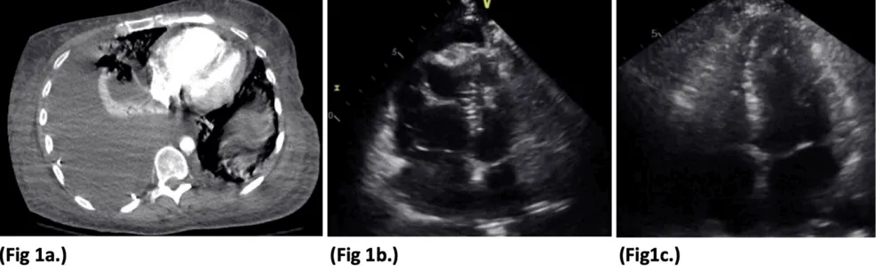
Selenium deficiency is a rare but reversible cause of cardiomyopathy increasingly recognized as a late nutritional complication of bariatric surgery, yet it is not consistently included in routine postoperative surveillance. This report describes a 40-year-old woman who presented three years after Roux-en-Y gastric bypass (RYGB) with progressive dyspnea, weakness, lower extremity edema, and dizziness in the setting of nonadherence to postoperative nutritional supplementation. She was hypotensive and bradycardic with jugular venous distension, elevated B-type natriuretic peptide (BNP), and chest imaging showing cardiomegaly with a right-sided pleural effusion. Echocardiography revealed severe, isolated right ventricular and atrial dilation with reduced right-sided systolic function, and further evaluation identified a markedly low serum selenium level (25 µg/L) with normal thyroid function and other trace elements. She was treated with intravenous selenium repletion (150 µg/day) alongside inotropic and diuretic support, with rapid hemodynamic improvement and complete resolution of right ventricular and atrial dilation on repeat echocardiography within one week. This case illustrates
Severe Right Ventricular Dilation Due to Selenium Deficiency in a Patient After Gastric Bypass - Cureus
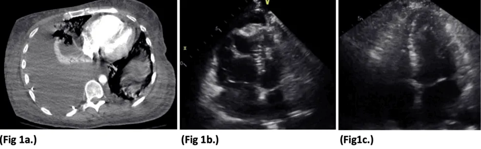
Selenium deficiency is a rare but reversible cause of cardiomyopathy increasingly recognized as a late nutritional complication of bariatric surgery, yet it is not consistently included in routine postoperative surveillance. This report describes a 40-year-old woman who presented three years after Roux-en-Y gastric bypass (RYGB) with progressive dyspnea, weakness, lower extremity edema, and dizziness in the setting of nonadherence to postoperative nutritional supplementation. She was hypotensive and bradycardic with jugular venous distension, elevated B-type natriuretic peptide (BNP), and chest imaging showing cardiomegaly with a right-sided pleural effusion. Echocardiography revealed severe, isolated right ventricular and atrial dilation with reduced right-sided systolic function, and further evaluation identified a markedly low serum selenium level (25 µg/L) with normal thyroid function and other trace elements. She was treated with intravenous selenium repletion (150 µg/day) alongside inotropic and diuretic support, with rapid hemodynamic improvement and complete resolution of right ventricular and atrial dilation on repeat echocardiography within one week. This case illustrates
Radiologically Suspected Ventricular Infection in a Patient With Recent Self-Extraction of a Carious Tooth: A Case Report - Cureus
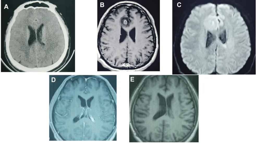
A brain abscess is a potentially life-threatening intracranial infection that usually requires individualized management. The possible source of infection may remain unclear when microbiological confirmation is unavailable. A 48-year-old man developed headache, fever, chills, and agitation approximately two weeks after self-extraction of a carious tooth. However, an odontogenic source was not microbiologically confirmed, and the temporal association alone did not establish causality. He also reported a history of swimming in a public pool, although the timing and characteristics of this exposure were unclear. After receiving empirical antimicrobial therapy at a local hospital without substantial improvement, he presented to Beijing Tiantan Hospital with a six-day history of fever and headache. Contrast-enhanced brain magnetic resonance imaging (MRI) showed a right frontal lesion radiologically consistent with a brain abscess and abnormalities of the left lateral ventricle and ependyma suggestive of ventriculitis, with possible intraventricular empyema. The suspected ventriculitis and intraventricular empyema were based on imaging findings and were not microbiologically confirmed. C
AI platform allows real-time conversation between patients and their medical devices - Medical Xpress
A new AI-driven platform could allow people to interact with their medical devices by sending messages using a smartphone application and receiving feedback in real time. Participants in a proof-of-concept study undergoing monitoring for seizures could ask questions about their brain activity, receive personalized alerts, report symptoms and behaviors and help train the system to better recognize seizure-related patterns.
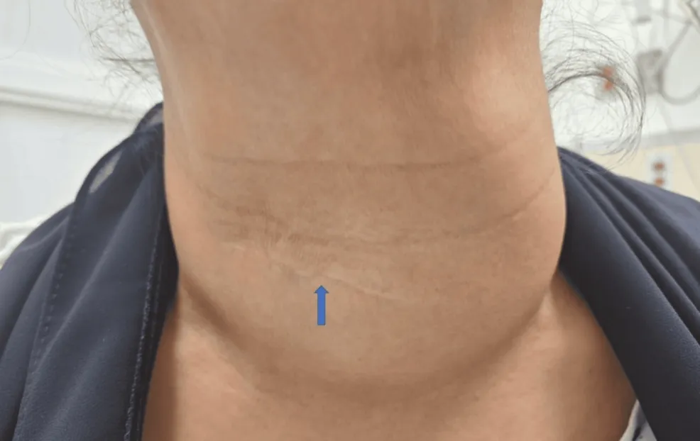
Acute Intrathyroidal Hematoma in a Multinodular Thyroid Presenting With Sudden Neck Swelling and Tracheal Deviation: A Case Report - Cureus
Acute hemorrhage within the thyroid gland is uncommon but can cause rapid neck swelling and, in severe cases, airway compromise. We report a 65-year-old woman with hypertension and a history of partial thyroidectomy performed more than 25 years earlier who presented with painful neck swelling that developed overnight. The swelling was associated with pain on swallowing, without shortness of breath, voice change, drooling, fever, or recent neck trauma. She was hemodynamically stable with an oxygen saturation of 99% on room air. Examination showed marked neck swelling, thyromegaly, thyroid tenderness, and left-sided neck induration. Contrast-enhanced computed tomography (CT) demonstrated a multinodular thyroid with a large heterogeneous mass in the left thyroid lobe causing mass effect and rightward tracheal deviation. Subsequent ultrasonography demonstrated a heterogeneous intrathyroidal collection consistent with hematoma. The patient remained clinically stable and was discharged after approximately five hours of observation. At otolaryngology follow-up 10 days later, repeat ultrasonography showed a progressively enlarging left thyroid nodule, and fine-needle aspiration cytology wa
0 comments
No comments yet. Be the first to share your thoughts!