Scientists uncover fossilized blood vessels in 66-million-year-old dinosaur bone - Interesting Engineering
This story deserves to be shared
Send it to someone who'd care, or post it on your networks.
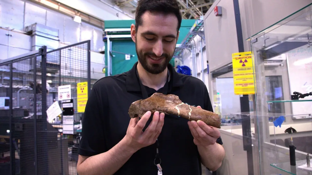
Scientists have unearthed rare fossilized blood vessels inside the broken rib of Scotty, the largest known Tyrannosaurus rex, using neutron imaging to peek inside the 66-million-year-old bone without damaging it.
The discovery provides a detailed view into how the dinosaur’s body responded to injury before its death and demonstrates how neutron imaging can reveal soft tissues that are rarely preserved in fossils.
This article is an excerpt. To read the full content, visit the original source. Interesting Engineering
Related articles
These are the first feathers ever found in fossilized dinosaur poop - Science News
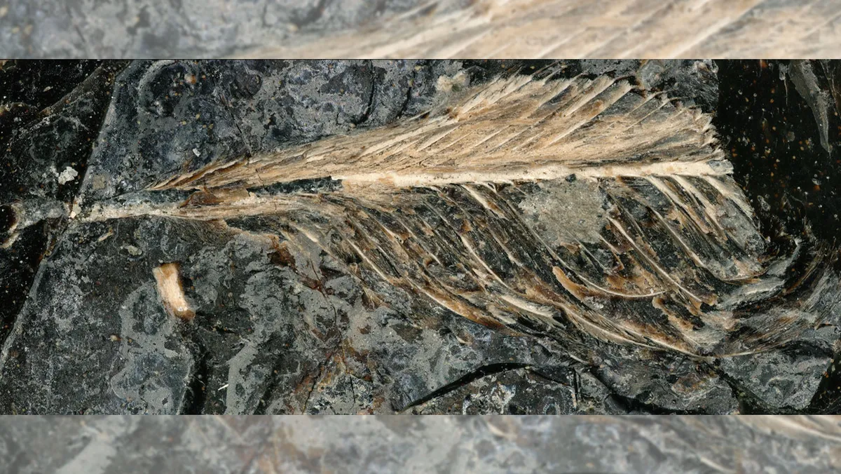
The plumage may help explain why only some birds survived dino doomsday Feathers like this one (shown at 200x magnification) preserved in fossilized dung from a meat-eating dinosaur such as a juvenile T. rex or Nanotyrannus could help explain why only some birds went extinct at the end of the Cretaceous Period.
Fossilized tissue reveals largest known T. rex died from broken rib - Popular Science
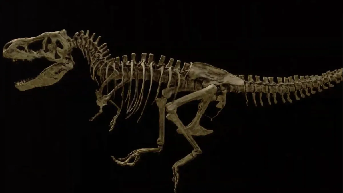
The largest Tyrannosaurus rex fossils ever discovered belong to a 66-million-year-old apex predator named Scotty. Like many T. rex skeletons, it’s difficult to determine if the specimen was male or female, but Scotty was an unequivocal bruiser. Standing 13-feet-tall at the hip and measuring upwards of 43-feet-long, the Late Cretaceous hunter weighed as much as 19,500 pounds. However, Scotty isn’t simply the biggest T. rex on record. One of its bones still has traces of preserved soft tissue. This is almost never seen in paleontology, as soft tissues degrade due to the ravages of time. The only way to safely examine these delicate remains is through highly sensitive techniques like neutron imaging. But with the help of researchers at the Department of Energy’s (DOE) Oak Ridge National Laboratory (ORNL) in Tennessee, paleontologists recently glimpsed Scotty’s rib and its remaining tissue in unprecedented detail. The results offer some of the first in-depth looks at fossilized dinosaur tissue on a microscopic level.
MIT Scientists Grew Artificial Blood Vessels In The Lab Using Magnets - ScienceAlert
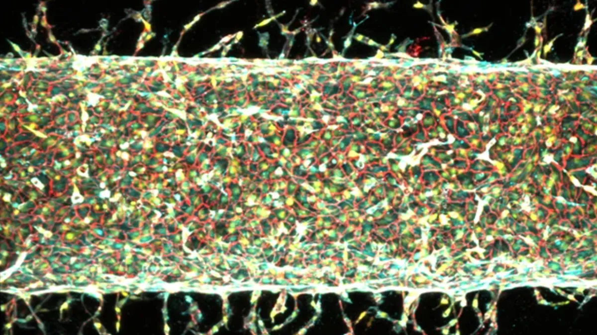
Science may one day give us a way to replace damaged and diseased parts of the body with artificial replacements – but reproducing organs and tissues in the lab isn't easy. That's especially true for networks of blood vessels, which at the level of fine, thread-like capillaries are microscopic – these capillaries can be as small as 0.005 millimeters (34 times thinner than a human hair), and only let blood cells through in single file. Researchers led by a team from MIT have now published a study in PNAS that details a way of engineering blood vessels in the lab with significantly greater precision than before. Getting the blood vessels (and therefore the blood flow) right is crucial to the success of any lab-grown organ or tissue, as fine capillaries perfuse tissue, delivering oxygen and nutrients. The artificial blood vessels were made through mechanical stretching. (MIT)The new approach is based on using magnetic forces that gently stretch and pull blood vessel cells into position.
0 comments
No comments yet. Be the first to share your thoughts!