New imaging technique detects subtle skin changes in scleroderma patients - News-Medical
This story deserves to be shared
Send it to someone who'd care, or post it on your networks.
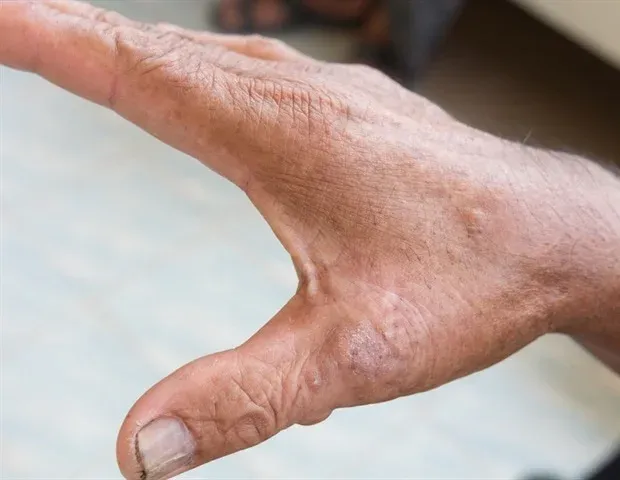
Scleroderma (systemic sclerosis) is a rare, chronic, autoimmune disease that causes the body to produce too much collagen, leading to the hardening and thickening of the skin and internal organs. For more than 50 years, physicians have relied on manually pinching the skin to assess its thickness, a subjective approach that remains central to the diagnosis of systemic sclerosis, assessment of disease extent and progression, and monitoring of response to treatment. Despite being the accepted standard practice, doctors can score the same patient differently based on their own unique touch and judgement, making it difficult to accurately track changes in skin thickening over time or assess a patient's response to treatment.
For the first time, researchers at Boston University Chobanian & Avedisian School of Medicine and the Boston University Department of Biomedical Engineering have developed and tested a camera-based technique called spatial frequency domain imaging (SFDI) to assess skin changes in scleroderma patients. SFDI works by shining patterns of harmless light on the skin, without touching it, and measuring how the light bounces back.
This article is an excerpt. To read the full content, visit the original source. News-Medical
Related articles

Scientists Puzzled as James Webb Detects Signal From Completely Empty Void in Space - Futurism
Using the NASA’s James Webb Space Telescope, a team of researchers led by University of Minnesota astrophysicist Mason Huberty had a closer look at a group of almost 10,000 galaxies spanning billions of light-years in the Fornax constellation. As detailed in a new paper published in The Astrophysical Journal, they honed in on a small space in the region that they thought was entirely unoccupied. But to their astonishment, they observed a single emission line emanating from it — twice over a span of just three months. Emission lines are bright, narrow bands of light emitted at a specific wavelength by distant celestial objects and other phenomena. Puzzlingly, prior observations from the same region didn’t feature the same reading.

Synchrotron imaging detects hidden lung risk for female farmers - Canadian Occupational Safety
A USask mouse study combining endotoxin and glyphosate exposure found lung changes only synchrotron imaging could detect A University of Saskatchewan (USask) study has found that combined exposure to two common agricultural contaminants – endotoxin and the herbicide glyphosate – produced lung damage in female mice that standard biomedical testing missed entirely, and that only became visible through synchrotron imaging at the Canadian Light Source (CLS) in Saskatoon. The research, led by Dr. Shelley Kirychuk, director of the Canadian Centre for Rural and Agricultural Health (CCRAH) at USask, was published this year in the journal Scientific Reports.
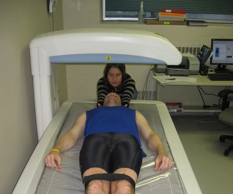
Bioengineer.org | Switching HIV drugs or adding zoledronic acid rebuilds bone lost to tenofovir - European AIDS Treatment Group
Bioengineer.org news storyFor millions of people living with HIV, antiretroviral therapy has transformed a once-fatal infection into a manageable chronic condition. Yet the very drugs that suppress the virus can carry quiet costs, and one of the most insidious is bone loss. Tenofovir disoproxil fumarate, or TDF, has been a cornerstone of HIV treatment worldwide for two decades, prized for its potency, durability, and low cost. But a growing body of evidence has linked TDF exposure to measurable reductions in bone mineral density, raising an increasingly urgent clinical question: once bone has been lost on a TDF-based regimen, can it be recovered? A new prospective cohort study, published in BMC Infectious Diseases, offers some of the most detailed longitudinal evidence to date that the answer, for many patients, is yes. The research team enrolled 223 virologically suppressed men living with HIV who had low bone mineral density while taking TDF-based antiretroviral therapy.
0 comments
No comments yet. Be the first to share your thoughts!